Miniature brain, eye, heart, kidney, artery, pancreas, neuron, and foot models. Education card illustrates the effects associated with Type II Diabetes: stroke, ocular pathology, hypertensive heart disease, hardening of the...
This model is designed as a visual learning aid for the musculature of the human head and neck, and the superior thoracic region. • This model represents the superficial and deep...
This 4-part model, 5X life-size, shows the human diencephalon: all the main parts of thalamus, epithalamus, metathalamus and hypothalamus are represented in great detail. The hypothalamic nuclei are displayed in...
This life-size model depicts 3 different pathologies that involve the vascular system of the brain, including arteriovenous malformations (called AVM), in which the blood vessels are tangled together rather than...
An important support for understanding the deeper anatomy of the brain. This single-piece model, enlarged 3 times, displays all the neurological tracts, exiting cranial and peripheral nerves in fine detail....
4x life size. Dissectible into 2 parts. The human cerebellum is dissected to show details of internal organization. Not on base. Includes numbered key. Every Dr Wong Anatomy® Model...
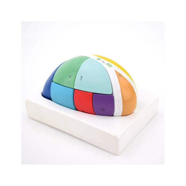
5X life-size. This 7-part model shows the various functional areas of the thalamus in distinct colors, thus providing a clear distinction between the different nuclei of each structure. Mounted on...
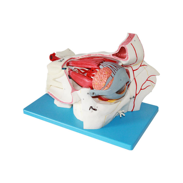
Experience an up-close and detailed view of the eye with this enlarged model. The frontal and sphenoid bones have been removed to showcase the bony orbit. With the ability to...
This life-size model is an important tool to study the topography of the cerebral ventricles related to the basal nuclei. All the different structures are reproduced in great detail and...