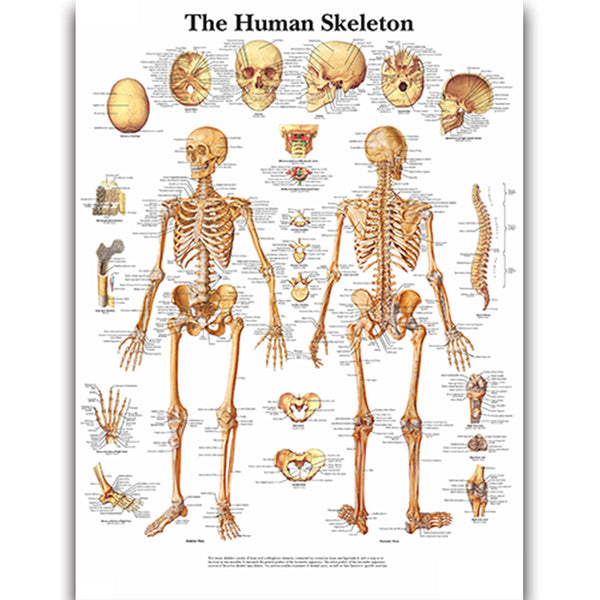
This detailed anatomical chart showcases the various components of the human skeleton. The skeletal anatomy is clearly labeled on a lifelike image, facilitating a comprehensive study of the body's bones....
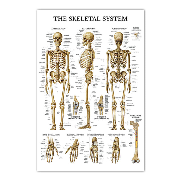
This detailed anatomical chart showcases the various components of the human skeleton. The skeletal anatomy is clearly labeled on a lifelike image, facilitating a comprehensive study of the body's bones....
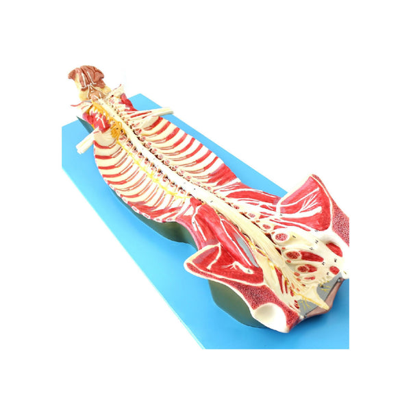
Seen from the ventral side, natural size. The model shows the brain stem and the spinal cord, as well as the nerve branches, up to the coccygeal plexus. On the...
This extremely detailed bone structure model depicts a three-dimensional section of a lamellar bone, showing a typical structure of a tubular bone enlarged 80 times. The bone structure model shows...
This model is composed of 4 parts and shows different structures of the human bones. The external structure of the femur with periosteum, blood vessels, spongy bone, compact bone, and...
This highly detailed model is designed as a visual learning aid for anatomy and physiology courses. Represents the human life-size cervical vertebral column with neck artery, occipital plate, herniated disc,...
This life size degenerative lumbar vertebrae disk disease model is designed as a visual leaning aid suitable for anatomy and physiologycourses, professional orthopedics, rheumatologists, chiropractic medicine, general anatomical study or rehab...
This highly detailed model is designed as a visual learning aid for anatomy and physiology courses. • Represents a cervical spine model with muscles showing the brain stem, occipital bone,...
This disarticulated skeleton model is designed to be used by any student of anatomy from beginner to advanced, and can be used by medical professionals, teachers and artists. • This...