This life-size model shows a frontal section of the human anus. Structures of the rectum, including the internal and external sphincter muscles, mucous membrane, ampullae, and anal valves are readily...
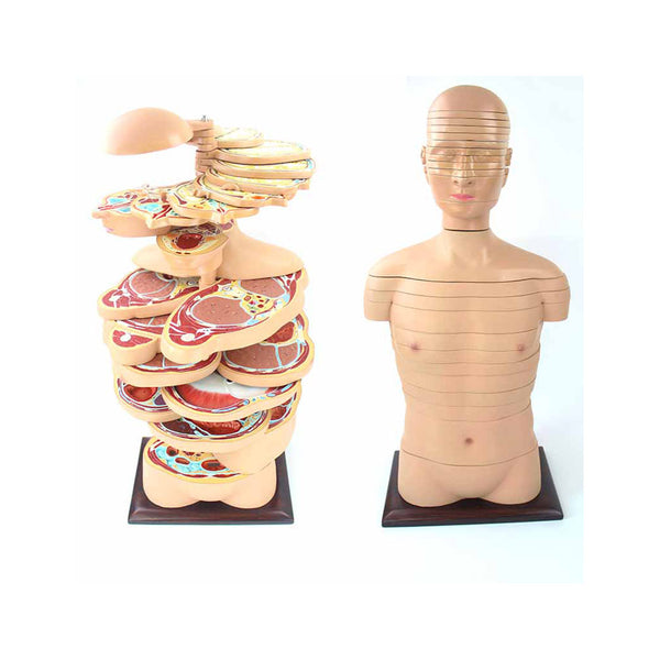
This life-size torso model is sliced horizontally into 24 pieces, giving an idea of how computer tomography and magnetic resonance work. Each slice can be rotated and removed for closer...
The Structure of the Stomach Wall Model is peeled off in a step-by-step manner to show the structure of each layer and the distribution of blood vessels, mainly showing the...
This intestinal villi model consists of one entire villus, one longitudinally sectioned villus showing the arterioles and venules and one sectioned villus to show the lymphatic vessels. The intestinal villi...
Enlarged approximately 400 times. The digitiform protrusions represent villi, the indentations show crypts. A cut surface reveals the histological structure of a villus. Cannot be disassembled. On a white base....
This model shows common stomach and intestinal diseases, such as gastritis and enteritis. The stomach is detachable into two parts, and the model is 1/2 life-size. Every Dr Wong...
This model is 2X life-size; it shows the open caecum with the appendix, ileum, ilocaecal orifice, and valve. Blood vessels and lymph nodes are also represented. Mounted on board....
Enlarged many times. The formation and structure of layers are shown by a transverse and vertical section. Model in one piece. On a blue base plate. Every Dr Wong Anatomy®...
This stomach pathology model demonstrates various stages of gastritis from a light gastric ulcer to a perforation. The stomach section with esophagus and duodenum attachment in half life size shows...