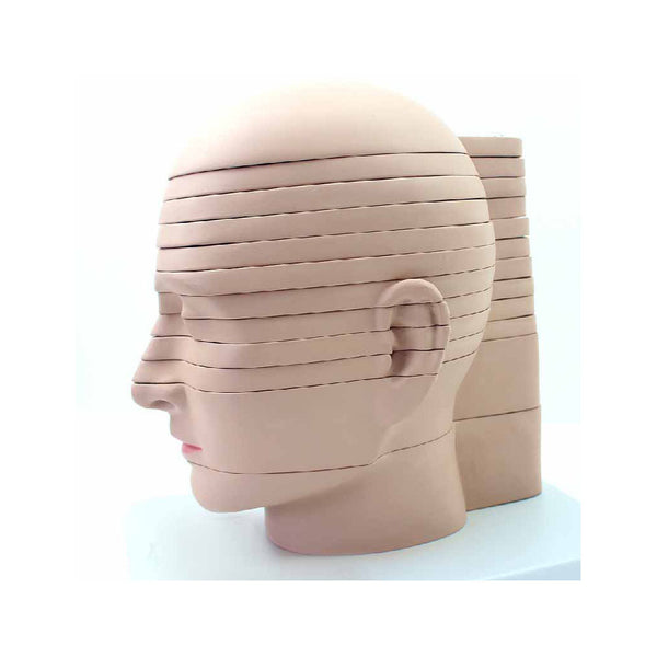
A meticulously hand-painted, sectioned model of the head is flawlessly detailed. This life-size head model is sliced horizontally into 12 pieces, giving an idea of how computer tomography and magnetic...
Enlarged model with removable parts for detailed anatomical study. ▪ 4X life size. ▪ The model shows the external, middle, and inner ear. ▪ The eardrum with malleus, incus, and stapes are removable....
▪ 3X life size. ▪ The model shows details of the external, middle, and inner ear. ▪ The eardrum with malleus, incus, and stapes are removable. ▪ The other removable part is composed of...
Enlarged approximately 4 times. Separates into pinna, petrous bone (3 parts), tympanic membrane with malleus and incus, labyrinth (2 parts), Eustachian tube. 8 parts in total. On a stand with blue...
Enlarged approximately 18 times. The network of nerves of the organ of balance is represented. Labyrinth Model mounted on a stand with a blue base. The Labyrinth Model is separated...
Enlarged approx. 19 times. Consisting of the malleus, incus and stapes. Model separates into 3 parts. On a stand with blue base. Every Dr Wong Anatomy® Model comes with a 3-year...
This 2-sided, average size, canine ear model features: a normal side with cochlea auditory tube tympanic bulla middle ear cavity tympanic membrane horizontal canal vertical canal auricular cartilage pinna and temporalis...
This single-piece model is a useful tool to study the mechanism of sound reception. The model shows an enlargement section of the cochlea and Corti’s organ including hair cells, tectorial...
Enlarged model of inner ear labyrinth, 2 parts This model is an enlarged model of the inner ear labyrinth. The model consists of two parts: an inner ear labyrinth (including...