This anatomical knee joint model accurately depicts the structure and functional movements of the human knee joint. It showcases internal and external rotation, flexion, extension, and abduction. The bones are...
This life size degenerative knee joint disease model is designed as a visual leaning aid suitable for anatomy and physiology courses, professional orthopedics, rheumatologists, chiropractic medicine, general anatomical study or rehab...
Human knee joint in life size with all important muscles and ligaments (collateral ligaments, meniscus, crucial ligaments, patellar tendon). The joint is not movable. On Stand. Every Dr Wong Anatomy®...
This 1/2 life-size model compares the normal knee joint with 3 different stages of ischemic pathology, from osteoporosis and atrophy to joint degeneration arthritis. Mounted on base Every Dr...
Life-size canine knee model with femur, fibula, patella, and tibia bones; lateral and medial meniscus; anterior and posterior cruciate ligaments; plus six more ligaments and tendons. Ideal for demonstrating veterinary anatomy. Every...
This model clearly shows the muscles, bones, vasculature, ligaments, tendons, and dermis presented in one convenient model. The knee joint is removable. Perfect for students, doctors, physical therapists, and more...
This anatomy model demonstrates the mechanism of meniscal tears. The different conditions that can be examined on the meniscus injuries model include: flap tear horizontal tear radial tear bucket handle longitudinal...
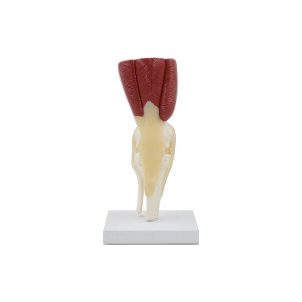
Full size normal right knee model includes: femur, fibula, patella and tibia bones; lateral and medial meniscus; quadriceps femoris tendon; anterior cruciate, fibular and tibial collateral, patellar and posterior meniscofemoral...
This is a life-size knee model that shows the location and function of the knee endoprosthesis. The prosthesis can be removed or replaced from the model, allowing the clinician to...